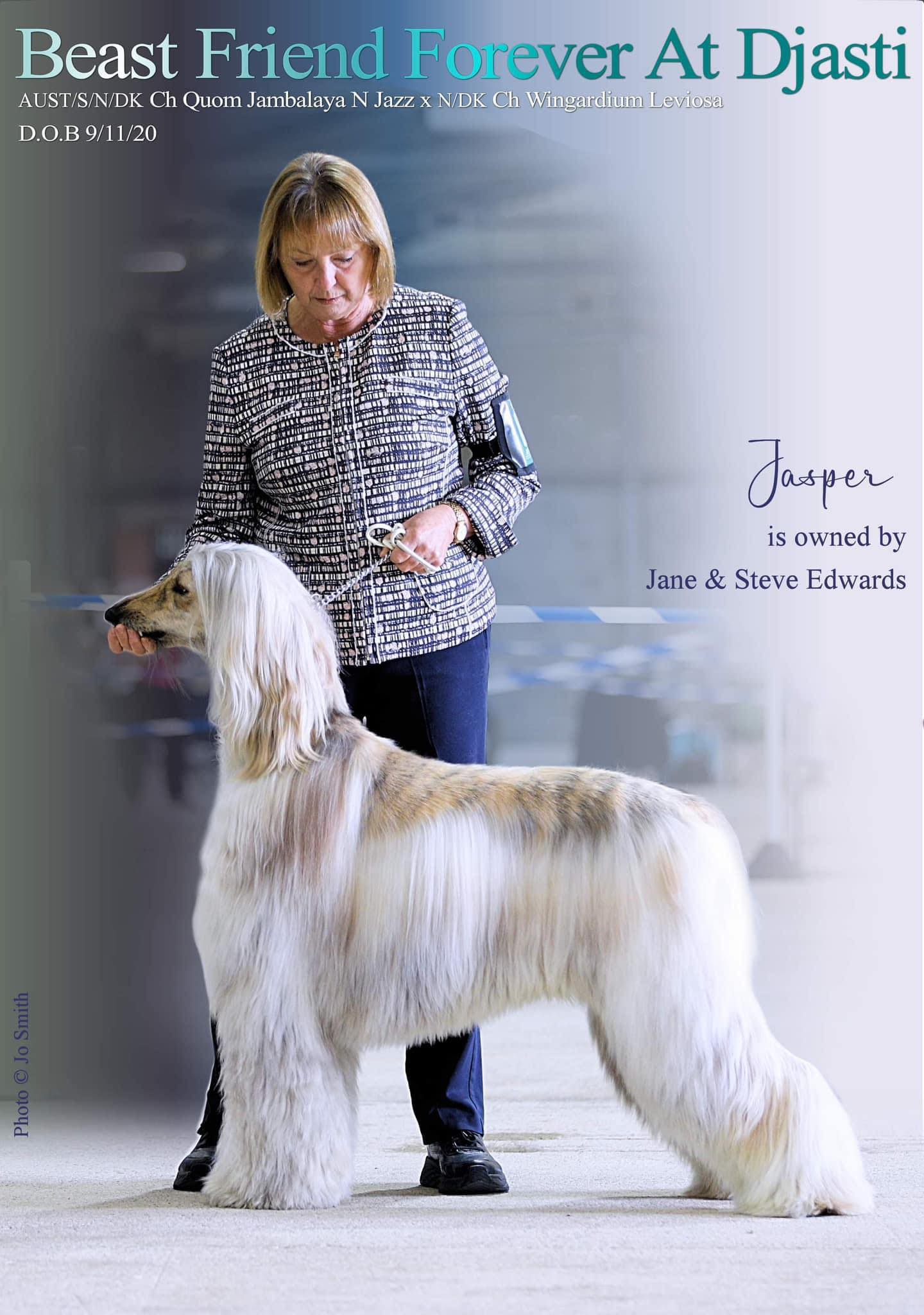
One of my Ringcraft Rebels show handling students has very recently gone through a terrifying ordeal. We are, fingers crossed, hoping they are now on the road to recovery but she desperately wanted to share her story with you and to lift the lid a little on a condition called Lung Lobe Torsion. Thank you Jane Edwards and her gorgeous boy Jasper for your time and consideration in making sure that we have as much knowledge of this condition as possible. Jane writes: ….
“I would like to make you aware of two rather nasty diseases that may darken your beautiful dog’s life. To be forewarned with some information could save their lives.
When is a Cough Not Kennel Cough ??
Lung Lobe Torsion:
Here are some symptoms to be aware of :- Lethargy, anorexia, coughing (which can sound like a goose honk) temperature/fever, a fast irregular breathing rate, the dog may start to suck in air, showing signs of stress, pale mucous membranes-which can turn blue, lips and gums etc.
Causes:- Spontaneous, idiopathic, previous surgery, secondary disease such as pneumonia, cancer.
Diagnosis is usually by one or more of these methods:-
X-ray, ultra sound, CT scan and or bronchoscopy.
Al dogs, all breeds and all sizes can be affected by this condition. Sight hounds unfortunately tend to be more predisposed to this condition than other breeds, due to their anatomical makeup, of being a large deep chested breed.
The dog’s lung lobes hang on “pedicles” off the Trachea, there are 3 on the Right hand side. At the top of the lung you have the largest lobe called the cranial, below is the middle/medial lobe with the bottom lobe being the caudal, there is also a small lobe which twists through from right to the left, called the accessory. On the Left hand side, there are 2, at the top is the cranial and below is the large caudal lobe. It is thought that due to the large deep chest of the sight hound, the lobes have more room in their thoracic cavity to move about. I believe in most sight hound cases, the right medial lobe is the one that twists.
.
In some dogs the condition may present with early clinical signs , in others it can be several months in duration.
.
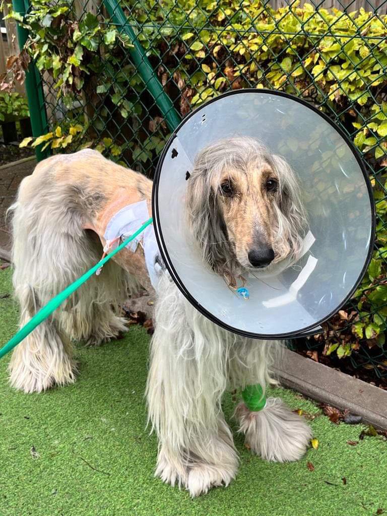
In our dogs case, he had no temperature and appeared well apart from a “soft” cough and slight lethargy. At first the cough was the main symptom, however Kennel Cough was doing its rounds, so we just kept him isolated and watched. After a couple of weeks there appeared to be no signs of the cough clearing up so we went to the vets and he was put on a course of antibiotics for 5 days. After the course ended he was still coughing but with a goose honk, still no fever. We took him back to the vets as he was beginning to show signs of respiratory distress. By the time we got him to our local vet hospital he was struggling to suck in air and his tongue was turning blue, he was very poorly. He was put on oxygen, and luckily a newly qualified vet, was studying our boys notes, and suggested to the team that it was LLT., Lung Lobe Torsion. they agreed, and to confirm diagnosis a CT scan was performed showing the build up of fluid in his abdominal cavity. A drain was placed in his right side, and 2 litres of pleural fluid was removed, this helped to stabilise him whilst an appointment was made to transfer him to a Specialist Referral Clinic.
.
On arrival at the Referral hospital we were met by a very professional veterinary nurse, who greeted and reassured us, whilst talking to us we were aware that she was gently going over him checking his clinical signs.. From a short distance we noticed our boys soft tissue surgeon observing how he was responding. The surgeon came over and took us to his consulting room. He explained that he had our boys notes from his vet and the CT scan, which confirmed the diagnosis. He then carefully explained the procedure of the operation. I asked about key hole surgery and he replied “No” due to the fact that he needs to remove the lobe intact as any spillage of necrotic fluid into the cavity may cause sepsis. The surgeon told us that they do about 2 LLT’s a week, scary!! It was explained that the surgery would take approximately 2:30hrs all going well. Once the surgery was over and our boy was in recovery in the ICU ward they would phone us for the results/outcome. Every second seemed like an hour, then the phone rang, the operation had gone according to plan, our boy was recovering, they had removed the lobe, stapled him together, put 2 drains in one on either side, put a drip/drain in all 4 legs, placed an oxygen tube up his nose, put him on a course of antibiotics, plus pain relief. Our poor, poor boy!
.
He remained in ICU for 2 weeks, during which we were given regular daily updates and photos which we waited eagerly for. As he progressed one drain was removed, the drips and oxygen and eventually , hurray! the final abdominal drain came out and he was allowed home with a list of Do’s and Don’ts, 3 x small meals per day and 3x short walks not exceeding 10 minutes per walk, and to watch his breathing rate , any concerns we were to ring them. Life was wonderful we were heading home!
Idiopathic Chylothorax:
.
I will now introduce you, unfortunately, to an even more devastating “rare” disease called in our case Idiopathic Chylothorax.
.
Symptoms: Lethargy, fever, respiratory issues, weight loss and a cough, in fact similar to LLT.
.
Our journey began approximately 3 weeks after coming home from his LLT operation, just when we thought all was going well, he started to cough….not much of a cough just a soft light cough once or twice, we thought that’s ok he is lying on his back, that’s what made him cough, out on a short walk I heard the “goose honk” I was getting paranoid, “he can’t be ill again!” He was eating well and before the LLT operation he had been in peak condition, which I am sure helped him with his recovery. I do confess I began researching with the aid of Dr Google! as I was becoming more concerned, and noted his breathing was becoming more rapid and he was panting ….showing signs of distress. So straight back to the local vets who agreed things were not good so an ultra sound was performed and yes fluid was building up. I add still a normal temperature, just the cough and rapid respiratory. Another 2 litres of fluid was drained out of him , as we waited for permission to be accepted back to his Specialist Clinic, I asked the vet who drained him if she thought the fluid was Chyle?? she replied that she had sent it off for analysis, but judging by the colour (creamy/ white) it looked that way. This was not what I wanted to hear.
.
I have enjoyed owning my breed for approx. 50 years, and although I have known of this disease, I have not physically had anything to do with it.
Once more devastated, we set off back to our specialist Soft Tissue Surgeon.
.
Chylothorax is said to be a rare, it is a devastating disease that will strike puppies and older dogs. Chyle flows within the lymphatic system, briefly “one” of the jobs the lymphatic system has to do is transport fats that have been absorbed by the gastrointestinal tract, in order for these fats to be transported in the fluid the small intestine converts them into fat with a coat of protein these molecules are called chylomicrons, this process enables the fats to be dissolved and carried through the lymphatic system to the cisterna chyli which is found in the abdominal cavity. It then flows into the thoracic duct and in turn empties into the major veins of the heart.
.
Our boy was to have three surgeries, 1. Thoracic Duct Ligation, 2. Pericardiectomy and 3. Cisterna Chyli Ablation. These three procedures would take approximately 5:30hrs all going well.
.
I cannot tell you how totally destroyed we felt, again our poor boy having to go through yet again another dreadful operation, he was only 2years and 8months old, we prayed he would fight it , he was strong we told ourselves, he’ll come through…. yet again we waited for the phone call, you can’t get on with a job, you can’t concentrate, all you can do is pace up and down, look at your watch, to see how many hours they were in surgery, then eventually what seemed like years but was in fact 5:30hrs, the phone call came through, he was back in recovery!! it was a difficult operation, but they had a full team available , soft tissue, cardio thoracic and anaesthetists, at one stage that sweet boys heart went out of rhythm and was massaged back, but he was still fighting, he was now in recovery in the ICU ward.
.
He had drips and drains wherever there was a space on his body, oxygen, and on ketamine pain relief. After a week, it was decided that if we were prepared to drain him ourselves at home, it would be best for him to come home with us. So once more we sterilised the family room that was our dog room, hibi scrubbed his bed his duvets, throws and vet beds plus his Meg Bed. We bought low to fat free raw diet, Rutin from Holland and Barratt, to help with fat dispersal, not proven but most people with their dogs suffering from this evil disease swear by it, plus some good healthy steamed vegetables to give him some carbohydrate, and a vitamin and mineral supplement. He was back on the 3x small meals a day plus the 3x 10minute walks. We had a lesson in how to drain him, which was to be once a day roughly at the same time each day. We were sent off with a bag full of medical swabs, syringes, dressings and various surgical gloves. He looked so very broken when we took him home, he was so very thin and just laid in his bed sleeping all day and night, only waking to eat his food which he wolfed down, he would not drink water from his water bowl, but I got water into him by making an island in his food bowl of his raw food and pouring water, just a little around the edges, so we had no problems with dehydration, I could not leave him at night, so I slept on the sofa next to him, sharing turns with my husband, day by day he slowly improved.
.
We began draining every day, some days draining 250mls then the next day it would be 180mls then it would bounce back to 200mls, it was so soul destroying to see it go back up, then it started to stay down and get less, our surgeon then told us to do it alternate days to see if we could kick start the body to drain/absorb the fluid itself, the fluid would at times be thinnish then it would change colours, from the creamish colour to coral and darker to a raspberry colour. After some time the volume began dropping steadily, then the drain stopped working, so we went back to the Clinic, where they removed the drain, and ultra sound him, they said we could take him home and watch him, as the drain may be causing him some irritation, so far so good. He has now been allowed free running which was a joy to see this beautiful sight hound running free once more….I add in an enclosed dog field. We live day to day hoping never to see the return of this devastating disease.
.
My intention was to write this blog in order that it might just help someone in need, and also from an owner’s simplistic knowledge and words of living with this wretched disease that so little is known about. I wish all your dogs a happy and healthy life.”
.
Thank you so much Jane for taking the time to share Jasper’s long, arduous and terrifying journey. He will ALWAYS be my little prince xx